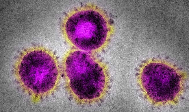
A critique of electron microscopy of "covid 19" for the purpose of identification.
Electromicrograms have been presented as COVID-19 by the NIH and others without question.
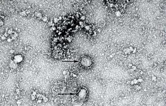
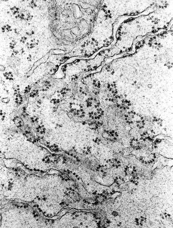
I will offer one simple example of the issue, first, you must look at two images of COVID-19.
Now, both of these are coronavirus? Incorrect. The last is the endoplasmic reticulum.
The CDC confirmed that the virus is ‘difficult’ to differentiate from organelles (components of the cell):https://wwwnc.cdc.gov/eid/article/27/4/20-4337_article
‘Difficulties in Differentiating Coronaviruses from Subcellular Structures in Human Tissues by Electron Microscopy’
The publication states
‘ribosomes along the endoplasmic reticulum may be confused with viral spikes. Ribosomes of vesiculating RER are in direct contact with the cell cytoplasm, unlike coronavirus spikes, which would be in contact with vacuolar contents. Vesiculating RER lacks cross sections through the viral nucleocapsid. Scale bar indicates 1 µm.’
If a person is actively searching for a structure, it is easy for a person to miscategorise the structure based on it having similar morphology. Take the example of mushrooms. Even the best mycologist struggles to tell certain species apart and will avoid them. The differences between the morphology of two mushrooms of the same species can be as different as two mushrooms of separate species. Now, Mushrooms have much more morphological characteristics than what is described as COVID-19.
The large endoplasmic riticulum has rings of ribosomes which are situatiated in such a way as to aid protein synthesis, this can give the false impression of a circular structure.
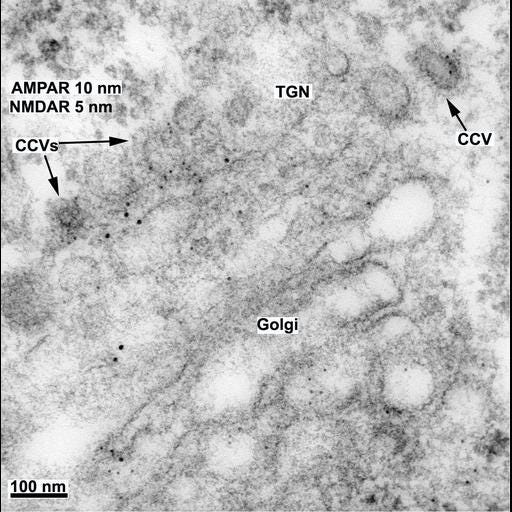
Other than this Clathrin-coated vesicles have also been noted as causing misidentification, with the protein coats being misinterperated as spike protein.
(CCV is clathrin coated vesicles)
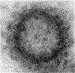
Now we will see how the images of covid-19 are presented to the public:
The image is coloured, outlining the percieved structure and making the ‘spikes’ stand out much more. The original image is in black and white and it has likely been edited in other ways.
Conclusion: electron micrographs are a poor test of the presence of viruses within tissues because the virus cannot be distinguished from the tissue itself. Until there is proof that the human eye can distinguish between viruses and biologicsl tissue the test cannot be considered scientific, the results of any such a test may be purely subjective in nature and could cause misidentification of illness. This could be a risk to human health.


